Brain Perfusion SPECT Scan in Hyderabad: Procedure, Uses, Cerebral Blood Flow and Neurological Assessment
By Focus Diagnostics
8 min read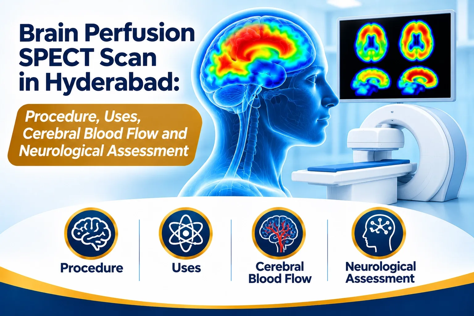
Brain Perfusion SPECT Scan in Hyderabad: Procedure, Uses, Cerebral Blood Flow and Neurological Assessment
The human brain requires a continuous supply of oxygen and nutrients through an extensive network of blood vessels. Even small changes in regional cerebral blood flow can affect brain function.
When doctors need to understand how blood is distributed across different regions of the brain, specialized functional imaging may be recommended.
A Brain Perfusion SPECT Scan is a nuclear medicine investigation that evaluates regional cerebral blood flow using a radioactive tracer and a specialized gamma camera.
Unlike routine structural imaging, which primarily demonstrates brain anatomy, perfusion SPECT provides functional information about blood flow patterns in different brain regions.
For patients searching for a brain perfusion SPECT scan in Hyderabad, understanding its clinical applications, imaging procedure and preparation can help them make informed decisions.
What Is a Brain Perfusion SPECT Scan?
Brain Perfusion Single Photon Emission Computed Tomography (SPECT) is a functional nuclear medicine imaging technique used to evaluate regional cerebral blood flow.
The examination commonly uses Technetium-99m-labelled radiopharmaceuticals such as:
- Tc-99m HMPAO (Hexamethylpropyleneamine Oxime).
- Tc-99m ECD (Ethyl Cysteinate Dimer).
These radiopharmaceuticals can cross the blood-brain barrier and distribute within brain tissue in relation to regional cerebral perfusion at the time of injection.
A gamma camera detects the emitted gamma radiation and reconstructs three-dimensional images of the brain.
The resulting images help specialists assess the distribution of cerebral blood flow and identify regional patterns of reduced or increased tracer uptake.
Why Do Doctors Recommend Brain Perfusion SPECT?
Brain perfusion SPECT may be considered when functional information about regional cerebral blood flow is required.
Selected clinical applications include:
1. Evaluation of Seizure Disorders
In selected epilepsy evaluations, brain perfusion SPECT may help identify regions associated with seizure activity.
Ictal SPECT, performed during or close to a seizure under a specialist-directed protocol, may demonstrate increased regional perfusion.
Comparison with interictal imaging can help localize a potential seizure focus during presurgical evaluation.
2. Assessment of Cerebrovascular Conditions
Perfusion imaging can provide information about regional cerebral blood flow in selected patients with cerebrovascular disorders.
It may be used in specialist-directed evaluations involving vascular insufficiency and selected cerebral perfusion abnormalities.
3. Evaluation of Dementia
Brain perfusion SPECT may demonstrate characteristic regional perfusion patterns in selected patients undergoing neurological assessment for cognitive decline.
These patterns can support clinical evaluation but cannot independently establish a dementia diagnosis.
4. Brain Death Evaluation
Specialized cerebral perfusion scintigraphy protocols may be used in selected circumstances as an ancillary investigation for assessing cerebral blood flow when determining death by neurological criteria.
This is a distinct, strictly protocol-governed examination and must be performed according to applicable clinical and legal standards.
5. Assessment of Cerebral Vascular Reserve
In selected patients with cerebrovascular disease, pharmacological challenge studies using agents such as acetazolamide may help evaluate cerebrovascular reserve.
This is a specialist-directed application rather than a routine brain perfusion study.
How Does Brain Perfusion SPECT Work?
Brain perfusion SPECT is based on the relationship between regional cerebral blood flow and the distribution of an appropriate radiopharmaceutical.
The imaging process generally involves the following stages:
Step 1: Clinical Assessment
The imaging team reviews the patient's medical history, symptoms, neurological evaluation and previous imaging reports.
The referring neurologist's specific clinical question helps determine the appropriate protocol.
Step 2: Patient Preparation
The patient is prepared in a quiet, controlled environment to minimize unnecessary sensory stimulation and changes in brain activity.
The preparation protocol depends on the indication.
Step 3: Radiotracer Administration
A small quantity of Tc-99m HMPAO or Tc-99m ECD is administered intravenously.
The tracer crosses the blood-brain barrier and becomes distributed in brain tissue in relation to regional perfusion at the time of injection.
Step 4: Uptake and Imaging
Following tracer administration, imaging is performed using a gamma camera.
The patient lies comfortably on the examination table while the camera acquires data from multiple angles.
Step 5: Image Reconstruction
The acquired information is processed to generate three-dimensional SPECT images of the brain.
Step 6: Specialist Interpretation
The nuclear medicine specialist evaluates regional tracer distribution, image quality and relevant clinical information before preparing the report.
What Are Tc-99m HMPAO and Tc-99m ECD?
Both radiopharmaceuticals are commonly used for cerebral perfusion SPECT.
Tc-99m HMPAO
HMPAO is a lipophilic radiopharmaceutical that crosses the blood-brain barrier and provides a representation of cerebral perfusion at the time of administration.
Tc-99m ECD
ECD is another lipophilic radiopharmaceutical used for regional cerebral perfusion imaging.
Both tracers help visualize the distribution of cerebral blood flow through nuclear medicine imaging.
The choice of tracer depends on clinical indication, availability and the department's validated imaging protocol.
What Does a Brain Perfusion SPECT Scan Show?
Brain perfusion SPECT provides functional information about tracer distribution in different regions of the brain.
A specialist may assess:
- Regional cerebral blood flow patterns.
- Areas showing relatively reduced tracer uptake.
- Areas showing relatively increased tracer uptake.
- Differences between ictal and interictal imaging when clinically indicated.
- Patterns relevant to selected neurological and cerebrovascular conditions.
A reduced uptake pattern is not specific to one disease. It must be interpreted in the context of clinical findings, structural imaging and other neurological investigations.
Brain Perfusion SPECT vs MRI Brain: What Is the Difference?
Both investigations provide valuable but different types of diagnostic information.
FeatureBrain Perfusion SPECTMRI BrainMain focusRegional cerebral perfusionBrain anatomy and tissue structureTechnologyNuclear medicine gamma cameraMagnetic resonanceRadiotracerYesNo in routine structural MRIFunctional informationRegional perfusion patternsDepends on MRI sequenceCommon applicationsSelected epilepsy and neurological evaluationsTumours, stroke, inflammation and structural abnormalities
The choice depends on the clinical question and the neurologist's recommendation.
Brain Perfusion SPECT vs CT Perfusion vs MRI Perfusion
These techniques assess cerebral perfusion using different technologies.
SPECT Perfusion: Uses radiopharmaceutical distribution to demonstrate regional cerebral blood flow.
CT Perfusion: Uses contrast-enhanced CT imaging to assess cerebral blood flow, blood volume and transit-related parameters.
MRI Perfusion: Uses specialized MR techniques to evaluate perfusion and related tissue characteristics.
Each modality has different strengths and limitations. The appropriate test depends on the clinical indication, urgency, patient suitability and availability.
How Should You Prepare for a Brain Perfusion SPECT Scan?
Preparation depends on the indication and the radiopharmaceutical protocol.
Patients may be advised to:
- Carry the neurologist's prescription and previous imaging reports.
- Inform the team about all current medications.
- Disclose a history of seizures or recent seizure episodes.
- Follow instructions regarding caffeine and other stimulants.
- Avoid unnecessary sensory stimulation before tracer administration.
- Inform the medical team about pregnancy or possible pregnancy.
- Discuss breastfeeding before the examination.
Patients should not independently stop anti-seizure medication or any other prescribed medicine.
The imaging department will provide specific preparation instructions according to the examination protocol.
Is a Brain Perfusion SPECT Scan Safe?
Brain perfusion SPECT is an established nuclear medicine investigation.
It uses a controlled quantity of a radioactive tracer, and radiation exposure depends on the administered activity and imaging protocol.
The examination is generally well tolerated.
Possible considerations include:
- Minor discomfort at the injection site.
- Radiation exposure from the radiopharmaceutical.
- Difficulty remaining still during imaging.
- Special precautions for pregnancy and breastfeeding.
For children and other special patient groups, radiopharmaceutical activity and imaging protocols must be appropriately adjusted.
Where Can You Enquire About Brain Perfusion SPECT in Hyderabad?
Brain perfusion SPECT is a specialized nuclear medicine investigation that requires appropriate radiopharmaceuticals, equipment and trained professionals.
Patients should confirm the availability of the exact examination, clinical protocol and appointment requirements before visiting a diagnostic facility.
Focus Diagnostics provides diagnostic services in Hyderabad.
Patients can contact the team to enquire about specialized imaging investigations and appointment coordination.
Phone / WhatsApp: 91213 77123
Please confirm the availability of brain perfusion SPECT before scheduling an appointment.
Frequently Asked Questions About Brain Perfusion SPECT
1. What does a brain perfusion SPECT scan detect?
It evaluates regional cerebral blood flow and can demonstrate patterns of reduced or increased tracer uptake associated with selected neurological and cerebrovascular conditions.
2. Is brain perfusion SPECT used for epilepsy?
Yes. In selected patients, particularly during presurgical epilepsy evaluation, ictal and interictal SPECT imaging can help identify regions associated with seizure activity.
3. Which radiotracer is used for brain perfusion SPECT?
Commonly used radiopharmaceuticals include Technetium-99m HMPAO and Technetium-99m ECD.
4. What is the difference between MRI brain and brain perfusion SPECT?
MRI primarily evaluates brain anatomy and tissue characteristics, while perfusion SPECT evaluates regional cerebral blood flow using a nuclear medicine tracer.
5. Where can I enquire about a brain perfusion SPECT scan in Hyderabad?
Contact Focus Diagnostics at 91213 77123 or visitto enquire about the availability of specialized nuclear medicine investigations.
Conclusion
Brain Perfusion SPECT is a specialized nuclear medicine imaging technique that provides valuable functional information about regional cerebral blood flow.
It has selected applications in epilepsy evaluation, cerebrovascular assessment and neurological investigations where understanding cerebral perfusion is clinically important.
For patients in Hyderabad, understanding this advanced imaging technique can support informed discussions with neurologists and nuclear medicine specialists.
Focus Diagnostics – Supporting Better Healthcare Decisions Through Reliable Diagnostic Services.
Medical disclaimer: This article is intended for general educational purposes and does not replace consultation with a qualified neurologist or nuclear medicine specialist. The decision to perform brain perfusion SPECT should be based on individual clinical evaluation and specialist recommendations..