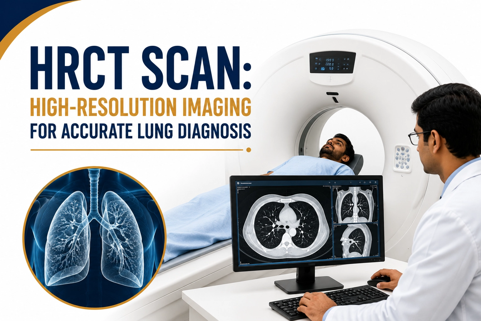
Brain SPECT Scan in Hyderabad: Uses, Procedure, Preparation and Results
By Focus Diagnostics
7 min readWhat Is a Brain SPECT Scan?
A brain SPECT scan, or brain single-photon emission computed tomography, is a nuclear medicine imaging test that provides information about the distribution of a radiotracer in the brain. A small amount of radiopharmaceutical is administered, and a gamma camera rotates around the head to collect data that are reconstructed into three-dimensional images.
When a perfusion tracer is used, the study can provide information about relative blood flow patterns in different regions of the brain at the time the tracer is administered. It is a functional imaging test and provides different information from structural imaging examinations such as CT or MRI.
Why Might a Doctor Recommend Brain SPECT?
A brain SPECT study may be considered in selected clinical situations when the treating specialist believes that functional information could contribute to the assessment. The exact indication and value of the test depend on the clinical question, the tracer and the imaging protocol.
Depending on the indication, a specialist may consider brain SPECT as part of an evaluation involving:
- Selected neurological or cerebrovascular conditions where regional cerebral perfusion information is required.
- Assessment of certain seizure-related questions, when appropriate to the clinical history and timing of imaging.
- Selected cases where functional information is sought in addition to structural imaging and other investigations.
- Specific specialist-led evaluations in which the result is expected to influence clinical assessment or management.
Brain SPECT is not a routine screening test for every headache, memory complaint or neurological symptom. Your neurologist or treating specialist should explain the reason for the test and how the result may affect your care.
How Does Brain Perfusion SPECT Work?
For a cerebral perfusion study, a radiotracer is administered through a vein. The tracer distribution reflects aspects of cerebral blood flow around the time of injection. After an uptake period, images are acquired using a rotating gamma camera. The scanner detects radiation emitted by the tracer and reconstructs a three-dimensional representation of its distribution.
Some protocols include CT images for attenuation correction or anatomical reference. The exact tracer, timing, acquisition method and image processing depend on the clinical indication and local protocol.
How to Prepare for a Brain SPECT Scan
Preparation can vary considerably according to the reason for the scan and the tracer being used. Follow the instructions provided by your treating specialist and imaging team. Before your appointment:
- Share a list of all prescription medicines, over-the-counter medicines and supplements you take.
- Tell the team about seizure history, recent seizures, neurological symptoms and relevant previous diagnoses.
- Bring previous CT, MRI, EEG and other relevant reports or images if available.
- Inform the team if you are pregnant, might be pregnant or are breastfeeding.
- Tell the staff if you have difficulty lying still, significant anxiety or any condition that may affect the imaging procedure.
- Ask whether food, drink, caffeine, medicines or activity need to be restricted for your particular protocol.
Do not stop prescribed medicines or change your usual treatment unless your treating doctor specifically instructs you to. Some studies require careful control of the conditions before tracer injection, so accurate preparation information is important.
What Happens During the Procedure?
- Clinical review: The team confirms the referral, reviews relevant history and explains the steps of the examination.
- Preparation: You may be asked to rest in a quiet environment before tracer administration, depending on the protocol.
- Tracer administration: A small amount of radiopharmaceutical is usually injected into a vein. The team will tell you when the tracer is administered and what to expect.
- Uptake period: You may need to wait while the tracer distributes. Instructions may include remaining calm and avoiding unnecessary movement or stimulation.
- Image acquisition: You lie on the imaging table with your head positioned carefully. The gamma camera rotates around your head while images are collected. It is important to remain still.
- Completion: The images are checked for technical quality. The team will let you know if additional images are needed and provide any post-scan instructions.
The total appointment time varies according to the tracer, uptake period and imaging protocol. Confirm the expected duration with the centre before your visit.
Is a Brain SPECT Scan Safe?
Brain SPECT uses a small, carefully selected amount of radiopharmaceutical. The protocol is designed to obtain the required diagnostic information while keeping radiation exposure as low as reasonably achievable. The tracer is usually administered through a vein, so brief discomfort at the injection site may occur.
Tell the team before tracer administration if you are pregnant, might be pregnant or are breastfeeding. The appropriateness of the examination and any precautions depend on the radiopharmaceutical and clinical circumstances. Your treating specialist can discuss the expected benefits and potential risks in your individual situation.
Understanding Brain SPECT Results
A nuclear medicine physician interprets the images in the context of the referral and available clinical information. The report may describe the distribution of tracer uptake and whether there are areas of relatively increased or reduced uptake compared with other regions.
A perfusion pattern is not a diagnosis by itself. Results can be influenced by the patient's condition at the time of tracer injection, technical factors, movement and the specific protocol. The interpreting physician and treating specialist consider the findings alongside symptoms, examination, MRI or CT, EEG, laboratory tests and other relevant information.
Do not start, stop or change treatment based solely on an imaging report. Discuss the findings and their relevance to your care with the specialist who requested the scan.
Brain SPECT vs CT and MRI
CT and MRI primarily provide structural information about the brain, although specialised MRI techniques can also provide functional or physiological information. Brain perfusion SPECT provides a map of radiotracer distribution that can reflect regional perfusion at the time of tracer administration. These tests answer different questions and may be complementary in selected cases. The treating specialist decides which investigation is appropriate.
Brain SPECT Scan in Hyderabad
If your doctor has recommended a brain SPECT scan in Hyderabad, ask which clinical question the examination is intended to address, which tracer and protocol will be used, and what preparation is required. Bring relevant previous reports and confirm the expected appointment duration. Focus Diagnostics can be contacted for information about the appointment process and current availability of the requested nuclear medicine study.
Phone: 91213 77123
Website: focusdiagnostics.in
Please confirm directly with the centre whether this specific examination is currently available before visiting.
Frequently Asked Questions
1. What does a brain SPECT scan show?
It shows the distribution of a radiotracer in the brain. With a perfusion tracer, the images provide information about relative regional blood flow around the time of tracer administration.
2. Is a brain SPECT scan the same as a brain MRI?
No. MRI primarily provides structural information, while brain perfusion SPECT provides functional information about tracer distribution. The tests may answer different clinical questions.
3. Does a brain SPECT scan diagnose every neurological condition?
No. Its usefulness depends on the clinical question and protocol. It is not a universal screening or diagnostic test for all neurological symptoms.
4. Do I need to fast before a brain SPECT scan?
Preparation depends on the tracer and clinical protocol. Follow the specific food and drink instructions given by your imaging team.
5. Should I stop my regular medicines?
Do not stop prescribed medicines on your own. Share your medication list and follow instructions from your treating specialist and imaging team.
6. Is the scan painful?
The imaging itself is non-invasive. A needle is usually used to administer the tracer, which may cause brief discomfort.
7. How long does a brain SPECT scan take?
The duration varies with the tracer, uptake period and image acquisition protocol. Ask the centre for the expected appointment time.
8. Can I have a brain SPECT scan if I am pregnant or breastfeeding?
Inform the team before tracer administration. They will assess whether the study is appropriate and provide advice specific to the radiopharmaceutical.
9. Should I bring my MRI or CT reports?
Yes. Bring relevant previous brain imaging, EEG reports and clinical records if available. They may help the interpreting physician understand the clinical context.
10. Where can I enquire about a brain SPECT scan in Hyderabad?
Contact Focus Diagnostics at 91213 77123 or visit focusdiagnostics.in to ask about the requested study, preparation and current availability.
Important Medical Note
This article is for general educational purposes only and does not replace medical advice, diagnosis or treatment. The need for brain SPECT imaging and the choice of tracer and protocol must be determined by a qualified treating specialist and nuclear medicine team.