Doppler Ultrasound Hyderabad: Types, Uses, Procedure & Preparation
By Focus Diagnostics
8 min read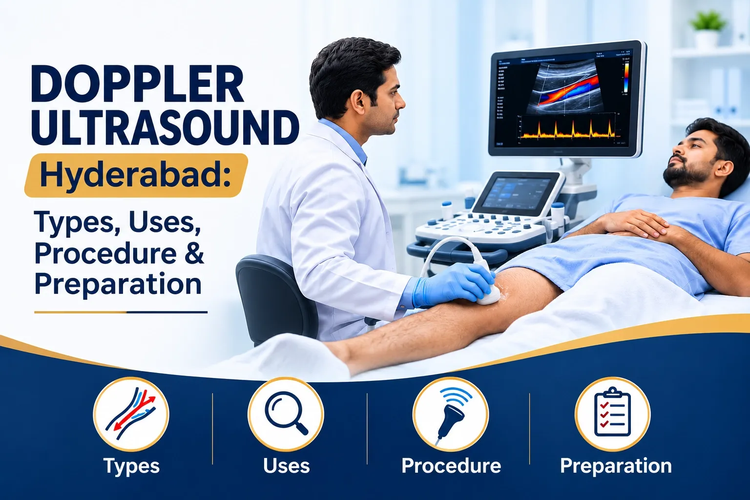
Doppler Ultrasound Scan in Hyderabad: Types, Uses, Procedure & Preparation
Understanding Doppler Ultrasound
Blood circulation plays a vital role in delivering oxygen and nutrients throughout the body. A Doppler Ultrasound Scan is a specialised imaging examination that helps evaluate the direction and speed of blood flow through selected blood vessels.
Unlike a conventional ultrasound, which primarily shows the structure of organs and tissues, Doppler imaging provides additional information about blood circulation, flow patterns and possible vascular abnormalities. Doctors may recommend this examination when investigating circulation-related symptoms or monitoring specific medical conditions.
For patients searching for a Doppler Ultrasound Scan in Hyderabad, understanding the purpose, types and preparation can help make the diagnostic process easier.
Early evaluation of abnormal blood flow can help doctors identify conditions that may require further investigation or treatment.
What Is a Doppler Ultrasound Scan?
A Doppler ultrasound uses high-frequency sound waves to assess blood movement through arteries and veins. The reflected sound waves help generate information about blood-flow direction and velocity.
This examination may be used to evaluate circulation in the limbs, neck, abdomen and selected blood vessels during pregnancy, depending on the doctor's recommendation.
Key features of Doppler Ultrasound:
- Evaluates blood flow through selected blood vessels.
- Helps investigate suspected narrowing or blockage.
- Supports assessment of certain blood clots and circulation problems.
- Uses sound waves instead of ionising radiation.
- Is generally non-invasive and painless.
Why Do Doctors Recommend a Doppler Scan?
A Doppler ultrasound may be recommended when symptoms or clinical findings suggest an abnormality in blood circulation.
Common reasons include:
- Unexplained swelling, pain or tenderness in a leg.
- Suspected deep vein thrombosis (DVT).
- Reduced blood circulation in the arms or legs.
- Suspected narrowing of selected arteries.
- Evaluation of blood flow in certain abdominal vessels.
- Assessment of selected fetal and maternal blood-flow patterns during pregnancy.
The type of Doppler examination depends on the symptoms, medical history and area that needs evaluation.
Types of Doppler Ultrasound Scans
Arterial Doppler Ultrasound
Arteries carry blood from the heart to different parts of the body. An arterial Doppler evaluates blood flow through selected arteries and may help identify reduced circulation or abnormal flow patterns.
Doctors may recommend this examination for symptoms such as leg pain while walking, cold extremities or suspected peripheral arterial disease.
Venous Doppler Ultrasound
Veins carry blood back towards the heart. A venous Doppler assesses blood flow through selected veins and is commonly used when a blood clot is suspected.
A leg Venous Doppler may help evaluate:
- Unexplained leg swelling.
- Pain or tenderness in the affected limb.
- Suspected deep vein thrombosis.
- Selected venous circulation abnormalities.
Color Doppler Ultrasound
Color Doppler displays blood-flow information as colour overlays on the ultrasound image. These colours help the radiologist assess flow direction and patterns according to the machine's settings.
It is often combined with conventional ultrasound and other Doppler techniques for a more comprehensive examination.
Carotid Doppler Ultrasound
The carotid arteries are located in the neck and supply blood to the brain. A carotid Doppler may be recommended to evaluate suspected narrowing or other abnormalities affecting these vessels.
Abdominal Doppler Ultrasound
An abdominal Doppler evaluates selected blood vessels supplying abdominal organs, including vessels associated with the liver and kidneys, when clinically indicated.
The exact examination depends on the doctor's request and the suspected vascular condition.
Pregnancy Doppler Ultrasound
An obstetric Doppler evaluates selected blood-flow patterns involving maternal and fetal circulation.
Depending on the clinical indication, it may assess the umbilical artery or uterine arteries to help evaluate placental circulation and fetal well-being. The examination should be recommended and interpreted by the obstetric care team.
Common Conditions Evaluated with Doppler Ultrasound
Doppler imaging provides useful information when doctors investigate certain vascular conditions.
Conditions that may be evaluated include:
- Deep Vein Thrombosis (DVT): A blood clot within a deep vein.
- Peripheral Arterial Disease: Reduced blood supply to the limbs due to arterial narrowing or blockage.
- Venous Insufficiency: Impaired blood return through the veins, sometimes associated with swelling or varicose veins.
- Vascular Narrowing: Changes in vessel diameter that may affect blood flow.
- Selected Abdominal Vascular Conditions: Abnormalities affecting blood supply to certain organs.
- Pregnancy-Related Circulation Concerns: Selected fetal or maternal blood-flow abnormalities.
A Doppler scan is not a substitute for a complete clinical assessment. The findings must be interpreted alongside symptoms, medical history and other investigations.
Symptoms That May Require Medical Evaluation
Certain symptoms may prompt a doctor to consider Doppler imaging.
Possible warning signs include:
- Persistent or unexplained swelling in one leg.
- Leg pain associated with walking.
- Unusual coldness in an arm or leg.
- Changes in skin colour associated with circulation problems.
- Symptoms suggesting a blood clot.
These symptoms can have several causes, and not everyone with them requires a Doppler scan.
Important: Sudden chest pain, breathlessness or a suddenly cold, pale and severely painful limb requires urgent medical attention. Do not delay emergency care to arrange an outpatient scan.
How Is a Doppler Ultrasound Scan Performed?
A Doppler ultrasound is generally performed using an ultrasound probe and a water-based gel applied to the skin.
During the examination, the imaging professional moves the probe over the relevant area to visualise the blood vessels and assess blood-flow patterns.
What to expect during the scan:
- You may be asked to lie down or change position.
- Ultrasound gel is applied to the examination area.
- The probe is moved over the skin to capture images.
- Doppler techniques assess blood-flow direction and velocity.
- The images are reviewed to prepare the diagnostic report.
The examination is usually painless, although mild pressure may be felt during some vascular studies. The duration varies depending on the area being examined and the complexity of the scan.
How to Prepare for a Doppler Ultrasound
Preparation depends on the type of examination being performed. Many leg and arm Doppler scans require minimal preparation, while certain abdominal studies may require fasting.
Before your appointment:
- Follow the preparation instructions provided by the diagnostic centre.
- Confirm whether fasting is necessary.
- Wear comfortable clothing that allows access to the examination area.
- Carry your doctor's referral and previous scan reports.
- Inform the imaging team about relevant medical conditions and medications.
- Continue prescribed medication unless your doctor advises otherwise.
For pregnancy Doppler examinations, follow the instructions provided by your obstetrician.
Doppler Ultrasound vs Regular Ultrasound
Both examinations use sound waves, but they provide different types of information.
Regular Ultrasound
Primarily shows the structure, size and appearance of organs and tissues.
Doppler Ultrasound
Adds information about blood-flow direction, velocity and patterns in selected blood vessels.
Combined Ultrasound Examination
Conventional imaging and Doppler techniques may be used together when both anatomical information and blood-flow assessment are needed.
The appropriate examination depends on the clinical question and the doctor's recommendation.
Is Doppler Ultrasound Safe?
Doppler ultrasound uses sound waves and does not involve ionising radiation. It is generally considered safe when performed by trained professionals for appropriate medical indications.
The examination is non-invasive and usually does not require injections or contrast agents. Pregnancy-related Doppler studies should be performed when clinically indicated and according to professional guidance.
Understanding Your Doppler Scan Report
A Doppler report may describe the vessels examined, blood-flow patterns, measured velocities and any abnormalities identified during the examination.
Depending on the scan, the report may include:
- Blood-flow characteristics in the examined vessels.
- Evidence of suspected narrowing or obstruction.
- Findings related to venous blood clots.
- Relevant measurements and the radiologist's impression.
The significance of these findings depends on the complete report and your clinical condition. Your treating doctor can explain the results and advise whether further investigation or treatment is needed.
Doppler Ultrasound Scan in Hyderabad at Focus Diagnostics
If your doctor has recommended a Doppler Ultrasound Scan in Hyderabad, Focus Diagnostics can help you enquire about the relevant imaging service, appointment availability and preparation requirements.
Patients can contact the centre to confirm the availability of their required examination at a preferred location.
Focus Diagnostics locations:
- Punjagutta, Hyderabad
- Kompally, Hyderabad
- Khajaguda, Hyderabad
For appointment enquiries, call 91213 77123.
When contacting the centre, mention whether you require an arterial, venous, carotid, abdominal or pregnancy Doppler scan. Availability may vary by branch and examination type.
Frequently Asked Questions About Doppler Ultrasound
What is a Doppler ultrasound scan used for?
A Doppler ultrasound evaluates blood flow through selected blood vessels. It can help investigate suspected blood clots, reduced circulation and certain vascular abnormalities.
Is Color Doppler different from a normal ultrasound?
Yes. A conventional ultrasound primarily shows anatomical structures, while Color Doppler adds colour-coded information about blood flow.
Why is a Doppler scan recommended for the legs?
A leg Doppler may be recommended to investigate unexplained swelling, pain, suspected blood clots or reduced arterial circulation.
Is a Doppler ultrasound painful?
The examination is generally painless. Mild pressure may be applied with the probe during some vascular studies.
Do I need to fast before a Doppler scan?
Many peripheral vascular Doppler scans do not require fasting. Certain abdominal examinations may require it, so confirm the instructions before your appointment.
Is Doppler ultrasound safe during pregnancy?
Ultrasound does not use ionising radiation and is generally considered safe when medically indicated and performed appropriately. Pregnancy Doppler examinations should be guided by the obstetric care team.
Can Doppler ultrasound detect blood clots?
Venous Doppler ultrasound is commonly used to evaluate suspected deep vein thrombosis. Results must be interpreted alongside clinical findings.
How long does a Doppler ultrasound take?
The duration depends on the blood vessels being examined and the complexity of the study. Your diagnostic centre can provide an estimated appointment duration.
Do I need a doctor's referral for a Doppler scan?
Requirements vary by centre and examination type. A doctor's referral helps identify which vessels need assessment and which examination is appropriate.
Where can I enquire about a Doppler scan in Hyderabad?
You can contact Focus Diagnostics at 91213 77123 to enquire about scan availability, preparation requirements and appointment details.
Conclusion
A Doppler ultrasound scan helps evaluate blood circulation and supports the investigation of selected vascular and pregnancy-related conditions. Understanding the examination, its uses and preparation requirements can help patients approach their diagnostic appointment with confidence.