Lymphoscintigraphy Scan in Hyderabad: Procedure, Uses and Lymphatic System Imaging
By Focus Diagnostics
6 min read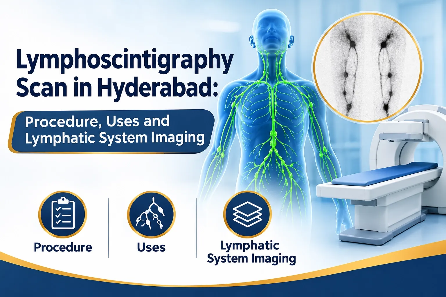
Lymphoscintigraphy Scan in Hyderabad: Procedure, Uses and Lymphatic System Imaging
The human lymphatic system plays an important role in maintaining fluid balance, supporting immune function and transporting lymph throughout the body. When lymphatic drainage becomes impaired, fluid can accumulate in tissues, potentially resulting in a condition called lymphedema.
But how do doctors evaluate the functioning of the lymphatic system?
Lymphoscintigraphy is a specialized nuclear medicine imaging investigation that helps evaluate lymphatic drainage and visualize the movement of lymphatic fluid towards regional lymph nodes.
For patients searching for a lymphoscintigraphy scan in Hyderabad, understanding the procedure, clinical applications and diagnostic importance can help them make informed decisions about their healthcare.
What Is Lymphoscintigraphy?
Lymphoscintigraphy is a functional nuclear medicine imaging technique used to evaluate the lymphatic system.
The examination involves administering a small quantity of a radioactive tracer, commonly a technetium-99m-labelled radiocolloid, into the tissues of the affected region.
The tracer travels through the lymphatic vessels and accumulates in regional lymph nodes.
A specialized gamma camera captures images over time, allowing nuclear medicine specialists to evaluate lymphatic drainage patterns.
Unlike conventional imaging techniques that primarily demonstrate anatomical structures, lymphoscintigraphy provides functional information about lymphatic transport.
Why Is a Lymphoscintigraphy Scan Recommended?
Doctors may recommend lymphoscintigraphy when they need to investigate abnormalities involving lymphatic circulation.
Common clinical applications include:
- Evaluation of lymphedema: Helps assess abnormal or delayed lymphatic drainage in patients with suspected primary or secondary lymphedema.
- Lymphatic drainage assessment: Demonstrates the movement of radiotracer through lymphatic channels towards regional lymph nodes.
- Sentinel lymph node mapping: Helps identify the first draining lymph node or nodes from a tumour site before selected cancer surgeries.
- Assessment of lymphatic abnormalities: Can demonstrate delayed transport, dermal backflow and abnormal drainage pathways.
- Preoperative planning: Provides functional information that may assist surgeons in selected lymphatic procedures.
The exact indication and imaging protocol depend on the patient's clinical condition.
How Does Lymphoscintigraphy Work?
Lymphoscintigraphy uses a radiopharmaceutical that enters the lymphatic circulation following administration into the appropriate tissue.
The procedure generally involves the following steps:
Step 1: Patient Assessment
The imaging team reviews the clinical history, symptoms, previous investigations and the doctor's referral.
Step 2: Radiotracer Administration
A small quantity of technetium-99m-labelled radiocolloid is administered through superficial injections, usually intradermally or subcutaneously, near the affected area or anatomical region being investigated.
Step 3: Dynamic Imaging
A gamma camera captures sequential images to observe the movement of the tracer through lymphatic channels.
Step 4: Delayed Imaging
Additional images may be acquired after a specified interval to evaluate tracer progression and regional lymph node visualization.
Step 5: Image Interpretation
The nuclear medicine specialist evaluates tracer movement, lymphatic drainage patterns, regional node uptake and any abnormalities.
The final report is shared with the treating doctor for clinical correlation.
Lymphedema is a condition characterized by swelling caused by the accumulation of protein-rich fluid in tissues due to impaired lymphatic transport.
It may develop due to congenital lymphatic abnormalities or damage to lymphatic vessels and nodes.
Common causes include:
- Previous cancer surgery involving lymph node removal.
- Radiation therapy.
- Congenital lymphatic abnormalities.
- Infection or injury affecting lymphatic structures.
- Certain conditions affecting lymphatic circulation.
Lymphoscintigraphy can demonstrate delayed lymphatic transport, abnormal tracer distribution and dermal backflow.
These findings may help doctors evaluate the functional status of the lymphatic system.
What Is Sentinel Lymph Node Mapping?
Sentinel lymph node mapping is an important application of nuclear medicine in selected cancer surgeries.
A sentinel lymph node is the first lymph node or group of nodes that receives lymphatic drainage from a particular tumour site.
In selected patients with breast cancer or melanoma, identifying these nodes helps surgeons determine which lymph nodes should be evaluated during surgery.
A radiotracer is injected near the tumour site, and imaging helps demonstrate the lymphatic drainage pathway and identify sentinel nodes.
The surgeon can subsequently use the mapping information to guide the procedure.
Sentinel lymph node mapping does not independently diagnose or exclude cancer in a lymph node. Tissue examination is required when histopathological confirmation is indicated.
Lymphoscintigraphy vs Ultrasound: What Is the Difference?
Both examinations can provide useful information, but they assess different aspects of the lymphatic system.
FeatureLymphoscintigraphyUltrasoundPrimary purposeFunctional lymphatic drainage assessmentAnatomical assessmentTechnologyNuclear medicine gamma cameraSound wavesTracer requiredYesNoLymphatic flow evaluationYesLimitedLymph node morphologyLimited anatomical detailDetailed structural assessmentCommon applicationsLymphedema and sentinel node mappingLymph node and soft-tissue assessment
The appropriate investigation depends on the suspected condition and the information required by the treating doctor.
How Should Patients Prepare for Lymphoscintigraphy?
Preparation requirements depend on the clinical indication and imaging protocol.
Patients should generally:
- Carry the doctor's prescription and previous imaging reports.
- Inform the imaging team about previous lymph node surgery.
- Disclose any history of cancer treatment or radiation therapy.
- Inform the medical team about pregnancy or possible pregnancy.
- Follow instructions regarding clothing and examination positioning.
Patients should confirm the preparation instructions with the imaging department before their appointment.
Is Lymphoscintigraphy Safe?
Lymphoscintigraphy is an established diagnostic nuclear medicine procedure that uses a small quantity of radiotracer.
Radiation exposure depends on the radiopharmaceutical and administered activity.
The examination is generally well tolerated. Minor discomfort may occur at the injection site.
Pregnancy and breastfeeding should be discussed with the nuclear medicine team before the procedure so that appropriate precautions and alternatives can be considered.
Where Can You Enquire About Nuclear Medicine Imaging in Hyderabad?
Patients who require specialized nuclear medicine investigations should understand the prescribed procedure and confirm its availability before scheduling an appointment.
Focus Diagnostics provides diagnostic services across Hyderabad, including locations such as Punjagutta, Kompally, KPHB, Madhinaguda, Chintal and Khajaguda.
Patients can contact Focus Diagnostics for information about diagnostic services, nuclear medicine investigation availability and appointment coordination.
Call / WhatsApp: 91213 77123
www.focusdaignostics.in
Frequently Asked Questions About Lymphoscintigraphy
1. What does a lymphoscintigraphy scan detect?
Lymphoscintigraphy evaluates lymphatic drainage and can demonstrate delayed tracer transport, abnormal lymphatic pathways and regional lymph node uptake.
2. Is lymphoscintigraphy used to diagnose lymphedema?
Yes. It can provide functional evidence of impaired lymphatic drainage and support the evaluation of suspected lymphedema.
3. Is lymphoscintigraphy used in cancer surgery?
Yes. In selected patients, it is used for sentinel lymph node mapping, particularly in certain breast cancer and melanoma procedures.
4. Is lymphoscintigraphy painful?
The imaging itself is painless. Patients may experience brief discomfort during the superficial radiotracer injections.
5. Where can I enquire about lymphoscintigraphy in Hyderabad?
Contact Focus Diagnostics at 91213 77123 to enquire about nuclear medicine services and confirm whether the specific investigation is available.
Conclusion
Lymphoscintigraphy is a specialized nuclear medicine investigation that provides valuable information about lymphatic circulation, drainage pathways and regional lymph node activity.
It plays an important role in selected cases of lymphedema evaluation and sentinel lymph node mapping.
For patients and doctors in Hyderabad, understanding this imaging technique can help support informed diagnostic decisions and appropriate clinical planning.
Focus Diagnostics – Supporting Better Healthcare Decisions Through Reliable Diagnostic Services.