Myocardial Perfusion Scan in Hyderabad: Procedure, Uses, Cardiac Blood Flow and Heart Disease Evaluation
By Focus Diagnostics
14 November 2026
8 min read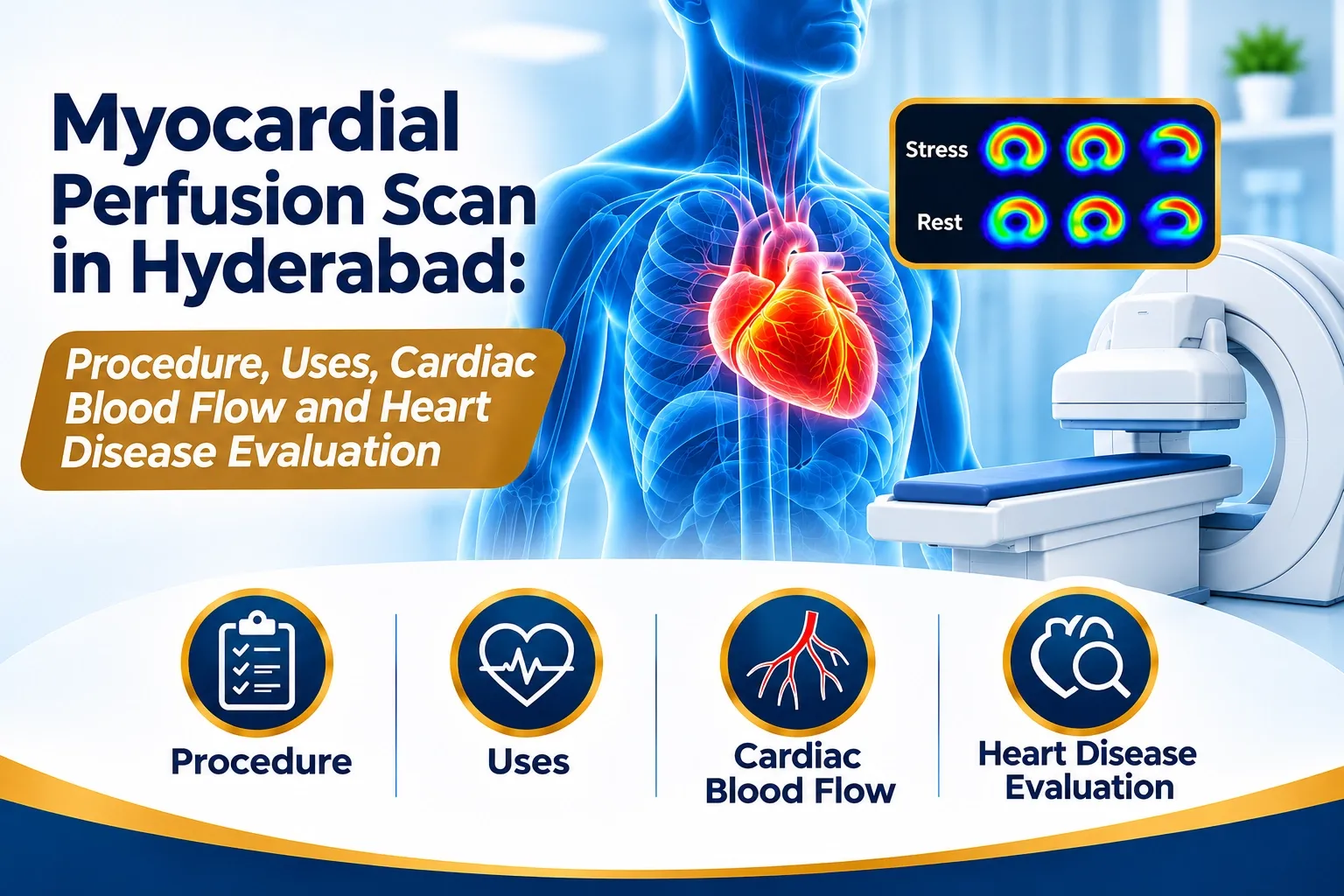
Myocardial Perfusion Scan in Hyderabad: Procedure, Uses, Cardiac Blood Flow and Heart Disease Evaluation
The heart continuously pumps blood throughout the body, supplying oxygen and essential nutrients to different organs. To perform this function effectively, the heart muscle itself requires an adequate supply of oxygen-rich blood through the coronary arteries.
When these arteries become narrowed or obstructed, the heart muscle may receive insufficient blood, particularly during physical activity or periods of increased cardiac demand.
A Myocardial Perfusion Scan (MPI) is a specialized nuclear medicine investigation that evaluates blood flow to the heart muscle under stress and resting conditions.
For patients searching for a myocardial perfusion scan in Hyderabad, understanding the procedure, clinical applications and diagnostic importance can help them make informed decisions about cardiac investigations.
What Is a Myocardial Perfusion Scan?
Myocardial Perfusion Imaging (MPI), also known as a nuclear cardiac stress test, is a diagnostic nuclear medicine examination used to assess blood supply to the heart muscle.
The examination uses a small quantity of a radioactive tracer, commonly Technetium-99m Sestamibi or Technetium-99m Tetrofosmin for SPECT imaging.
After administration, the tracer distributes within the heart muscle in relation to regional blood flow and cellular tracer uptake.
A specialized gamma camera captures images of the heart, allowing nuclear medicine specialists to evaluate the distribution of tracer activity.
The examination commonly includes stress and rest imaging to identify differences in myocardial perfusion.
Unlike a routine ECG, which records the electrical activity of the heart, MPI provides functional imaging information about blood flow to the heart muscle.
Why Do Doctors Recommend a Myocardial Perfusion Scan?
A cardiologist may recommend myocardial perfusion imaging when additional information about coronary blood flow or myocardial function is required.
Common clinical indications include:
- Suspected coronary artery disease: To assess whether narrowed coronary arteries may be limiting blood supply to the heart muscle.
- Evaluation of chest discomfort: To investigate selected patients with symptoms suggestive of myocardial ischemia.
- Previous heart attack: To evaluate areas of reduced perfusion that may be associated with previous myocardial injury.
- Assessment after angioplasty or bypass surgery: To evaluate myocardial perfusion in selected patients following coronary interventions.
- Cardiac risk assessment: To support risk assessment in appropriately selected patients with known or suspected coronary artery disease.
- Assessment of myocardial viability: In selected clinical situations, additional imaging and interpretation may help evaluate potentially viable heart muscle.
The decision to perform MPI should be based on clinical history, examination, previous investigations and the treating cardiologist's recommendation.
How Does Myocardial Perfusion Imaging Work?
Myocardial perfusion imaging evaluates how the heart muscle receives blood during different physiological conditions.
The procedure generally involves two imaging phases.
1. Stress Phase
The heart is placed under increased workload either through physical exercise or pharmacological stress.
During exercise stress, the patient may walk on a treadmill or use a stationary bicycle while heart rate, ECG and blood pressure are monitored.
If the patient cannot exercise adequately, an appropriate pharmacological stress agent may be administered under medical supervision.
A radioactive tracer is injected at the appropriate stage of stress to demonstrate myocardial perfusion under increased cardiac demand.
2. Rest Phase
Resting myocardial perfusion images are obtained using a tracer injection and imaging protocol appropriate to the examination.
The stress and rest images are compared to identify regional differences in tracer uptake.
3. Image Acquisition and Processing
A gamma camera captures the distribution of the radiotracer within the heart.
SPECT imaging reconstructs three-dimensional representations of myocardial tracer distribution.
Gated SPECT may additionally provide information about left ventricular wall motion, wall thickening and ejection fraction.
What Is the Difference Between Reversible and Fixed Perfusion Defects?
One of the important aspects of myocardial perfusion interpretation is comparing stress and rest images.
Reversible perfusion defect:
A region may show reduced tracer uptake during stress but relatively improved uptake during rest.
This pattern can suggest inducible myocardial ischemia, which means the heart muscle may not be receiving sufficient blood during increased demand.
Fixed perfusion defect:
A region may show reduced tracer uptake during both stress and rest.
This can be associated with previous myocardial infarction and scar tissue.
However, attenuation artifacts, technical factors and other conditions can also influence tracer distribution.
Interpretation must consider image quality, ECG findings, clinical history and other cardiac investigations.
What Is SPECT Myocardial Perfusion Imaging?
Single Photon Emission Computed Tomography (SPECT) is a nuclear medicine imaging technique commonly used for myocardial perfusion assessment.
In SPECT MPI, the radiotracer emits gamma photons that are detected by a rotating gamma camera.
The acquired data are processed to reconstruct three-dimensional images of the heart.
SPECT myocardial perfusion imaging can help evaluate:
- Regional myocardial blood flow.
- Stress-induced perfusion abnormalities.
- Fixed and reversible perfusion defects.
- Left ventricular ejection fraction through gated imaging.
- Regional myocardial wall motion and thickening.
SPECT-CT systems may also provide CT-based attenuation correction, helping reduce certain imaging artifacts.
What Is the Difference Between PET and SPECT Cardiac Imaging?
Both PET and SPECT are nuclear medicine imaging techniques, but they use different radiopharmaceuticals and imaging technologies.
FeatureSPECT MPIPET MPIImaging technologyGamma cameraPET scannerCommon tracersTc-99m Sestamibi or TetrofosminRb-82 or N-13 AmmoniaImaging outputMyocardial perfusion imagesMyocardial perfusion imagesAdditional assessmentGated cardiac functionCan quantify myocardial blood flow and flow reserveAvailabilityDepends on the imaging facilityDepends on specialized PET facilities
The choice depends on the clinical indication, available technology and cardiologist's recommendation.
How Should You Prepare for a Myocardial Perfusion Scan?
Preparation is important for obtaining diagnostically useful cardiac imaging.
Patients may be advised to:
- Avoid caffeine for the period specified by the nuclear medicine department, often 12–24 hours depending on the stress protocol.
- Follow instructions regarding fasting before the examination.
- Carry previous ECG, echocardiography, angiography and cardiac investigation reports.
- Inform the medical team about all prescribed medications.
- Discuss diabetes medications and insulin instructions before the appointment.
- Wear comfortable clothing and footwear if exercise stress is planned.
- Inform the medical team about asthma, breathing difficulties, pregnancy or possible pregnancy.
Important: Do not independently discontinue beta-blockers, nitrates or other cardiac medications. Medication adjustments must be directed by the treating cardiologist or nuclear medicine team.
Is a Myocardial Perfusion Scan Safe?
Myocardial perfusion imaging is an established diagnostic nuclear medicine procedure.
It uses a controlled quantity of radioactive tracer, and radiation exposure depends on the tracer, administered activity and imaging protocol.
Exercise or pharmacological stress testing also involves specific clinical considerations.
Possible risks include:
- Temporary fatigue or discomfort during exercise.
- Flushing, headache or breathlessness with certain pharmacological stress agents.
- Changes in heart rhythm or blood pressure.
- Rare serious cardiac complications during stress testing.
- Radiation exposure associated with the radiotracer.
The procedure is performed with appropriate medical supervision and monitoring.
Patients who are pregnant, may be pregnant or are breastfeeding should inform the treating team before the investigation.
Myocardial Perfusion Scan vs ECG vs Echocardiography
These cardiac investigations provide different types of information.
InvestigationMain purposeECGEvaluates electrical activity and heart rhythmEchocardiographyEvaluates heart structure, valves and pumping functionMyocardial Perfusion ScanEvaluates blood supply to the heart muscleCoronary CT AngiographyEvaluates coronary artery anatomyInvasive Coronary AngiographyDirectly evaluates coronary artery anatomy using catheter-based imaging
These tests are not interchangeable. Your cardiologist determines which investigation is appropriate for your clinical condition.
Where Can You Enquire About Cardiac Nuclear Medicine Imaging in Hyderabad?
Patients who have been advised to undergo myocardial perfusion imaging should confirm the exact test, stress protocol, preparation requirements and appointment availability before visiting a diagnostic centre.
Focus Diagnostics provides diagnostic imaging services in Hyderabad.
Patients can contact the team to enquire about cardiac imaging and nuclear medicine investigation availability, along with appointment coordination and preparation guidance.
Contact: Use the verified contact details listed on the official Focus Diagnostics website.
Frequently Asked Questions About Myocardial Perfusion Scan
1. What does a myocardial perfusion scan detect?
A myocardial perfusion scan evaluates blood flow to different regions of the heart muscle. It can help identify patterns suggestive of myocardial ischemia and previous myocardial injury.
2. Is a myocardial perfusion scan the same as a nuclear stress test?
Yes. Myocardial perfusion imaging is commonly called a nuclear stress test when stress and rest imaging are performed to evaluate cardiac blood flow.
3. How long does a myocardial perfusion scan take?
The total examination time varies according to the tracer, imaging protocol, stress method and whether rest and stress imaging are performed on the same day or different days.
4. Can a myocardial perfusion scan detect heart blockages?
It can identify patterns of reduced blood supply that may suggest functionally significant coronary artery disease. However, it does not directly visualize the coronary artery anatomy like coronary CT angiography or invasive coronary angiography.
5. Where can I enquire about a myocardial perfusion scan in Hyderabad?
You can contact Focus Diagnostics through its official website to enquire about cardiac nuclear medicine imaging availability, preparation instructions and appointment coordination.
Conclusion
Myocardial Perfusion Imaging is an important nuclear medicine investigation that helps evaluate blood supply to the heart muscle under stress and resting conditions.
By comparing myocardial perfusion patterns, doctors can assess possible ischemia, previous myocardial injury and selected aspects of cardiac function.
For patients in Hyderabad, understanding the role of cardiac nuclear medicine imaging can support informed discussions with their cardiologist and appropriate diagnostic planning.
Focus Diagnostics – Supporting Better Healthcare Decisions Through Reliable Diagnostic Services.
Medical disclaimer: This article is intended for general educational purposes and does not replace consultation with a qualified cardiologist or nuclear medicine specialist. The choice, protocol and interpretation of cardiac investigations should be based on individual clinical assessment.