Bone Scan in Hyderabad: Whole Body Bone Scintigraphy, Procedure, Uses and Cancer Detection
By Focus Diagnostics
8 min read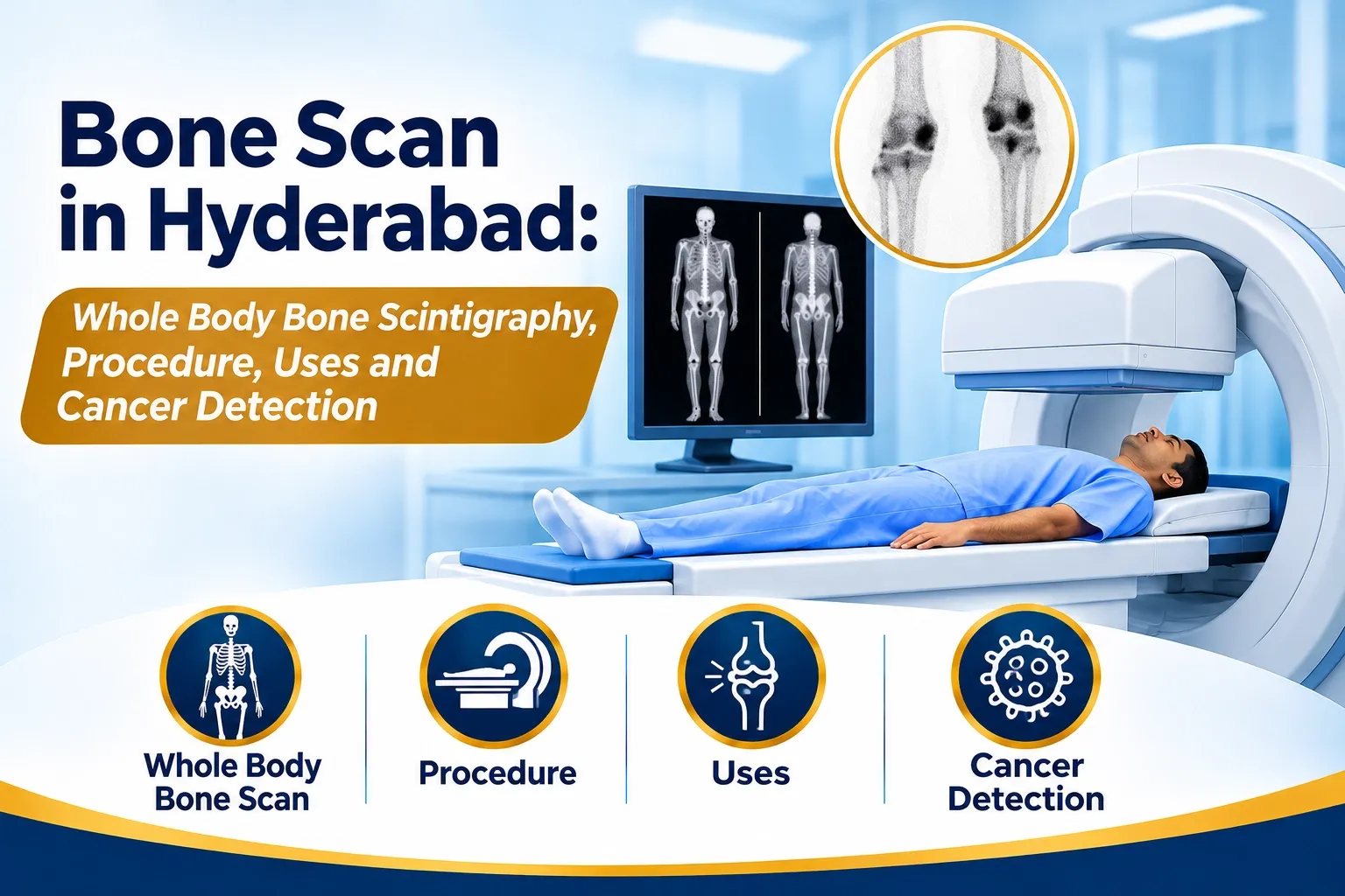
Bone Scan in Hyderabad: Whole Body Bone Scintigraphy, Procedure, Uses and Cancer Detection
Bones provide structural support, protect vital organs and enable movement. However, several medical conditions can affect bone metabolism, circulation, healing and overall skeletal health.
When doctors need to evaluate abnormal activity throughout the skeleton, a specialized nuclear medicine investigation called a Whole Body Bone Scan may be recommended.
Also known as bone scintigraphy or skeletal scintigraphy, this examination helps identify areas of increased or decreased bone turnover associated with certain cancers, fractures, infections and other skeletal conditions.
For patients searching for a bone scan in Hyderabad, understanding how this investigation works, what it detects and how to prepare can help make the diagnostic process easier.
What Is a Whole Body Bone Scan?
A whole body bone scan is a nuclear medicine imaging procedure used to evaluate metabolic activity throughout the skeleton.
The examination commonly uses a radioactive pharmaceutical called Technetium-99m Methylene Diphosphonate (Tc-99m MDP).
After intravenous injection, the radiotracer circulates through the bloodstream and accumulates in areas of bone formation and remodeling.
A specialized gamma camera detects the radiation emitted by the tracer and produces images of the skeleton.
These images help nuclear medicine specialists identify abnormal patterns of tracer distribution that may require further clinical evaluation.
Unlike conventional X-rays, which primarily demonstrate structural changes, bone scintigraphy provides information about skeletal metabolic activity.
Why Do Doctors Recommend a Bone Scan?
A doctor may recommend bone scintigraphy when there is a need to investigate suspected skeletal abnormalities or evaluate disease involving multiple bones.
Common clinical applications include:
1. Detection of Bone Metastases
In selected patients with cancer, a whole body bone scan can help identify skeletal abnormalities associated with metastatic disease, particularly osteoblastic or bone-forming responses.
It may be used in the evaluation of certain prostate, breast and lung cancers, depending on the clinical indication.
2. Investigation of Unexplained Bone Pain
Bone scintigraphy can help investigate persistent or unexplained bone pain when conventional imaging does not provide sufficient information.
3. Detection of Stress Fractures
It may help identify stress-related bone injuries and certain fractures that are not clearly visible on initial X-rays.
4. Evaluation of Bone Infections
In selected cases, bone scans may help investigate suspected osteomyelitis and other skeletal infections.
5. Assessment of Joint and Orthopaedic Conditions
Bone scintigraphy may provide useful information in selected patients with joint abnormalities, prosthetic joint complications and unexplained musculoskeletal symptoms.
6. Evaluation of Bone Remodeling Disorders
Certain metabolic and degenerative bone conditions can produce characteristic patterns of tracer uptake.
The final interpretation always depends on the patient's symptoms, medical history and other imaging findings.
How Does a Bone Scan Work?
Bone scintigraphy is based on the uptake of a bone-seeking radiopharmaceutical in areas of active bone remodeling.
The examination generally involves the following steps:
Step 1: Patient Assessment
The imaging team reviews the doctor's prescription, previous investigations, clinical history and relevant medical information.
Step 2: Radiotracer Injection
A small quantity of Tc-99m MDP is administered through an intravenous injection.
The tracer travels through the bloodstream and localizes in the skeleton according to regional bone turnover and blood supply.
Step 3: Uptake Period
The patient generally waits for approximately 2–4 hours, depending on the protocol.
During this period, adequate hydration and frequent urination are usually encouraged, unless medically restricted.
Step 4: Whole Body Imaging
The patient lies on the gamma camera examination table.
The camera moves over the body and captures anterior and posterior images of the skeleton.
Step 5: Image Interpretation
The nuclear medicine specialist examines tracer distribution and identifies areas showing abnormal uptake or other significant findings.
Additional spot views or SPECT/CT may be recommended when further anatomical localization is required.
What Is a Tc-99m MDP Bone Scan?
Technetium-99m Methylene Diphosphonate is one of the commonly used radiopharmaceuticals in conventional bone scintigraphy.
It is a bone-seeking compound that accumulates in areas of active mineralization and bone remodeling.
The distribution of Tc-99m MDP provides information about skeletal activity and helps identify abnormalities that may not yet produce obvious changes on conventional radiographs.
The tracer is detected using a gamma camera, and a computer processes the collected data into diagnostic images.
What Is the Difference Between a Three-Phase Bone Scan and a Whole Body Bone Scan?
Different bone scintigraphy protocols are selected depending on the suspected condition.
A three-phase bone scan includes three imaging stages:
Phase 1 – Flow Phase
Images are acquired immediately after tracer injection to evaluate blood flow to the region.
Phase 2 – Blood Pool Phase
Images are acquired shortly after injection to assess tracer distribution in the soft tissues surrounding the bones.
Phase 3 – Delayed Skeletal Phase
Images are obtained after the tracer has accumulated in the skeleton to evaluate bone uptake.
A conventional whole body bone scan primarily focuses on delayed skeletal imaging of the entire skeleton.
Three-phase imaging is particularly useful in selected evaluations involving infection, inflammation, painful prosthetic joints and other localized abnormalities.
How Can a Bone Scan Help Detect Cancer Spread to Bones?
Certain cancers can spread from their original location to the skeletal system. This process is known as bone metastasis.
Bone scintigraphy can help identify areas of abnormal bone remodeling associated with metastatic disease.
The examination is particularly useful for detecting many osteoblastic responses, although its sensitivity varies depending on tumour type and biological behaviour.
Areas of increased tracer uptake may appear as abnormal hot spots on the images.
However, increased uptake does not automatically mean cancer.
Fractures, arthritis, infections, inflammation and other benign conditions can also cause increased radiotracer accumulation.
Similarly, some predominantly osteolytic lesions may be less conspicuous on conventional bone scintigraphy.
Therefore, findings should be correlated with clinical information, CT, MRI, PET-CT or other appropriate investigations.
Bone Scan vs PET-CT vs MRI: What Is the Difference?
These imaging techniques provide different types of diagnostic information.
FeatureBone ScanPET-CTMRIMain focusSkeletal metabolic activityMolecular activity and anatomySoft tissue and bone marrow anatomyTechnologyGamma cameraPET and CTMagnetic resonanceCommon tracerTc-99m MDPDepends on PET tracerNo radioactive tracer in routine MRIWhole skeleton assessmentYesDepends on protocolUsually region-specificCommon applicationsBone metastases, fractures and selected infectionsCancer staging and selected bone diseaseBone marrow, soft tissue and musculoskeletal assessment
The most appropriate examination depends on the suspected disease, clinical question and treating doctor's recommendation.
What Does an Abnormal Bone Scan Mean?
An abnormal bone scan indicates an area of tracer distribution that differs from the expected pattern.
Possible explanations include:
- Bone metastases.
- Healing fractures.
- Degenerative joint disease.
- Bone infections.
- Inflammatory conditions.
- Metabolic bone disorders.
- Previous surgery or bone injury.
The pattern, location and intensity of tracer uptake are considered alongside the patient's medical history and other investigations.
An abnormal scan alone does not establish a specific diagnosis.
How Should You Prepare for a Whole Body Bone Scan?
Preparation for a conventional bone scan is generally straightforward.
Patients may be advised to:
- Carry the doctor's prescription and previous imaging reports.
- Inform the imaging team about recent fractures, surgery or bone injuries.
- Disclose any previous cancer diagnosis and treatment history.
- Inform the team about medications and relevant medical conditions.
- Drink adequate fluids after the radiotracer injection, unless fluid intake is restricted.
- Empty the bladder frequently and immediately before imaging.
- Inform the nuclear medicine team if pregnant, possibly pregnant or breastfeeding.
Fasting is generally not required for a routine bone scan, but the imaging department's specific instructions should always be followed.
Is a Whole Body Bone Scan Safe?
Bone scintigraphy is an established nuclear medicine diagnostic procedure.
It uses a small diagnostic quantity of a radioactive radiopharmaceutical.
The radiation exposure depends on the administered activity, tracer and imaging protocol.
The examination is generally well tolerated. Minor discomfort may occur during intravenous injection, and remaining still during imaging may be uncomfortable for patients with significant bone or joint pain.
Patients should inform the nuclear medicine team about pregnancy or breastfeeding before the examination so that the appropriate precautions can be considered.
Where Can You Enquire About Bone Imaging in Hyderabad?
Patients who have been advised to undergo a whole body bone scan should confirm the exact investigation, imaging protocol, preparation requirements and appointment availability before scheduling.
Focus Diagnostics provides diagnostic imaging services in Hyderabad.
Patients can contact Focus Diagnostics to enquire about available imaging investigations, appointment coordination and relevant diagnostic services.
Contact: 91213 77123
For specialized nuclear medicine investigations, please confirm service availability with the diagnostic team before visiting.
Frequently Asked Questions About Bone Scan in Hyderabad
1. What is a whole body bone scan used for?
A whole body bone scan evaluates skeletal metabolic activity and helps investigate conditions such as bone metastases, fractures, infections and selected bone disorders.
2. Can a bone scan detect cancer in the bones?
Yes. It can help identify skeletal abnormalities associated with cancer spread, particularly osteoblastic bone metastases. However, abnormal uptake is not specific to cancer and requires clinical correlation.
3. How long does a bone scan take?
The complete procedure commonly takes several hours because the tracer needs time to accumulate in the bones. Actual imaging time is generally shorter and depends on the protocol.
4. Is fasting required before a bone scan?
Fasting is generally not necessary for a routine bone scan. Patients are usually encouraged to maintain appropriate hydration and follow the department's preparation instructions.
5. Is a bone scan better than an MRI?
Neither investigation is universally better. Bone scintigraphy evaluates skeletal metabolic activity throughout the body, while MRI provides detailed anatomical information about bone marrow, soft tissues and specific body regions. The appropriate test depends on the clinical indication.
Conclusion
A Whole Body Bone Scan is an important nuclear medicine investigation that helps evaluate skeletal metabolic activity and identify abnormalities involving the bones.
From investigating unexplained bone pain and selected fractures to assessing possible cancer spread to the skeleton, bone scintigraphy provides valuable information for clinical decision-making.
For patients in Hyderabad, understanding the purpose, procedure and preparation of a bone scan can help support informed discussions with their treating doctors.
Focus Diagnostics – Supporting Better Healthcare Decisions Through Reliable Diagnostic Services.
Medical disclaimer: This article is intended for general educational purposes and does not replace consultation with a qualified medical professional. The choice and interpretation of nuclear medicine investigations should be guided by the treating doctor and nuclear medicine specialist.