Medically Reviewed By
Dr. Srinivas
Consultant Pathologist
Pathology · Last reviewed: June 2026
Book This Test
Fill in your details and connect with our team instantly on WhatsApp.
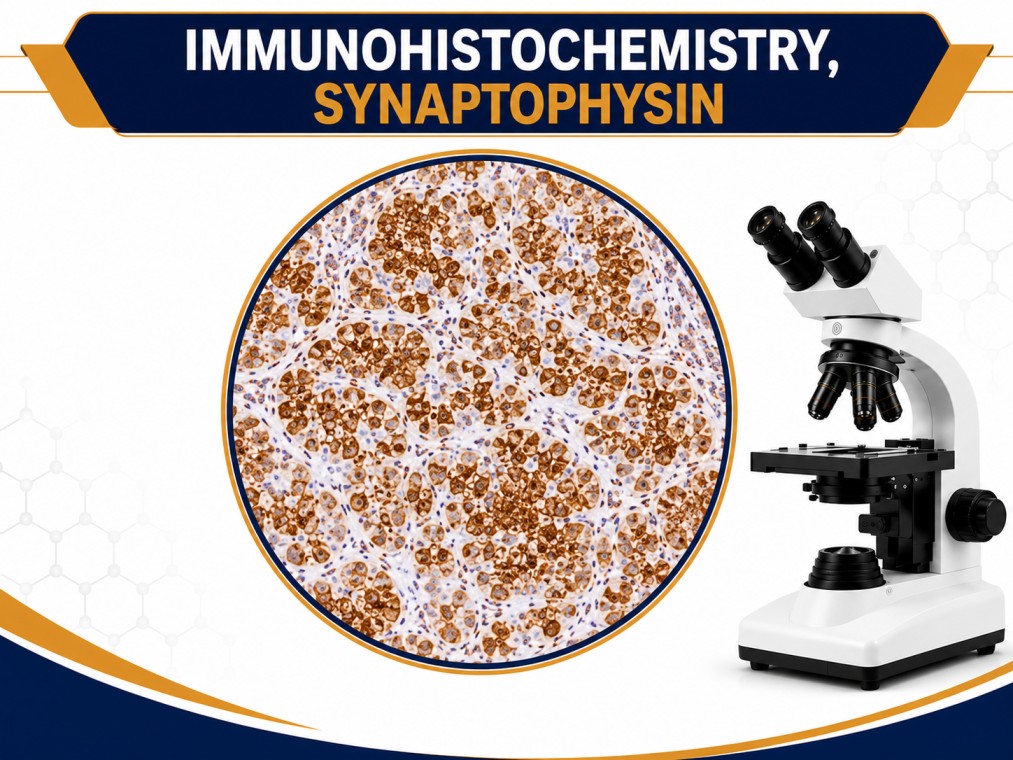
IMMUNOHISTOCHEMISTRY, SYNAPTOPHYSIN
Get reliable diagnostics, expert support, and a seamless booking experience with Focus Diagnostics.
About this test
The IHC – Synaptophysin test is an immunohistochemistry-based diagnostic assay used to detect Synaptophysin protein expression in tissue samples. Synaptophysin is a membrane glycoprotein found in presynaptic vesicles of neurons and neuroendocrine cells, making it one of the most important markers for identifying neuroendocrine differentiation in tumors.
This test is widely used in pathology and oncology to diagnose and classify neuroendocrine tumors (NETs). Synaptophysin staining helps pathologists determine whether tumor cells originate from neuroendocrine tissue and assists in distinguishing neuroendocrine tumors from other types of cancer.
The IHC Synaptophysin test is commonly used in combination with other neuroendocrine markers such as Chromogranin A, CD56, NSE, and INSM1 to improve diagnostic accuracy. It is considered one of the most sensitive markers for neuroendocrine differentiation and is frequently included in tumor immunohistochemistry panels.
Benefits of the Test
- Helps diagnose neuroendocrine tumors
- Assists in identifying neuroendocrine differentiation
- Supports tumor classification and grading
- Improves diagnostic accuracy in oncology
- Helps guide treatment planning and prognosis evaluation
Why Doctors Recommend This Test
Doctors recommend the IHC Synaptophysin test when evaluating suspected neuroendocrine tumors and related cancers. It is commonly used in:
- Neuroendocrine tumor diagnosis
- Carcinoid tumor evaluation
- Small cell carcinoma assessment
- Pancreatic neuroendocrine tumor investigation
- Lung neuroendocrine tumor classification
- Tumor pathology and oncology studies
Synaptophysin is often evaluated alongside Chromogranin A, CD56, Ki-67, Cytokeratins, and NSE to provide a comprehensive neuroendocrine tumor profile.
Preparation Before Test
No special preparation is required for this test. Important considerations include:
- The test is performed on a biopsy or surgical tissue sample
- No fasting is required
- No medication restrictions are generally necessary
- Clinical history and imaging findings may assist interpretation
Normal Reporting Time
The typical turnaround time is 3 to 5 working days, depending on tissue processing and laboratory workflow.
Who Should Take This Test
- Patients with suspected neuroendocrine tumors
- Individuals undergoing cancer biopsy evaluation
- Patients with small cell carcinoma
- Cases requiring tumor origin determination
- Oncology patients requiring immunohistochemistry profiling
The IHC Synaptophysin test is an essential diagnostic tool in modern pathology. By detecting neuroendocrine differentiation, it helps clinicians accurately diagnose tumors, classify cancer subtypes, and develop effective treatment strategies.
Clinical Significance
Synaptophysin immunostaining is commonly used in the evaluation of:
- Neuroendocrine tumors (NETs)
- Carcinoid tumors
- Small cell lung carcinoma
- Pancreatic neuroendocrine tumors
- Neuroblastoma
- Pheochromocytoma
- Paraganglioma
- Neuroendocrine carcinomas
Strong Synaptophysin positivity is a key indicator of neuroendocrine differentiation and plays a vital role in tumor classification.
Test FAQs
What is the IHC Synaptophysin test?
What does Synaptophysin positivity indicate?
Is this a blood test?
Which cancers are associated with Synaptophysin expression?
Why is Synaptophysin important in pathology?
How long does the test take?
Is fasting required?
What sample is needed?
Is Synaptophysin used alone for diagnosis?
Who interprets the report?
Find Your Nearest Focus Diagnostic Centre Hyderabad
Popular Lab Tests in Other Cities
Telangana
Andhra Pradesh
Popular Tests in Hyderabad
MRI
- MRI Brain
- MRI Brain with Contrast
- MRI Brain Angiography (MRA Brain)
- MRI Spine Cervical
- MRI Spine Dorsal
- MRI Spine Lumbar
- MRI Whole Spine Screening
- MRI Knee Joint
- MRI Shoulder Joint
- MRI Hip Joint
- MRI Ankle / Foot
- MRI Wrist / Hand
- MRI Pelvis
- MRI Abdomen
- MRI MRCP
- MRI Orbit
- MRI Internal Auditory Canal (IAC)
- MRI Neck
- MRI Breast
- MRI Whole Body Screening
CT-Scan
- CT Brain
- CT Brain with Contrast
- CT Angiography Brain
- CT Chest (HRCT Chest)
- CT Thorax
- CT Abdomen & Pelvis
- CT KUB (Kidney Ureter Bladder)
- CT Cervical Spine
- CT Dorsal Spine
- CT Lumbar Spine
- CT PNS (Sinus)
- CT Temporal Bone
- CT Neck
- CT Pulmonary Angiography (CTPA)
- CT Coronary Angiography
- CT Urogram
- CT Whole Abdomen
- CT Pelvis
- CT Facial Bones
- CT Lung Screening
Ultrasound – Special Scans
- Ultrasound Abdomen
- Ultrasound Pelvis
- Ultrasound Abdomen & Pelvis
- Ultrasound KUB
- Ultrasound Thyroid
- Ultrasound Breast
- Obstetric Ultrasound (Pregnancy Scan)
- NT Scan
- TIFFA / Level 2 Scan
- Growth Scan
- Fetal Wellbeing Scan
- Follicular Study
- Transvaginal Scan (TVS)
- Scrotal Ultrasound
- Ultrasound Soft Tissue
- Ultrasound Hernia
- Doppler – Carotid
- Doppler – Lower Limb Venous
- Doppler – Lower Limb Arterial
- Doppler – Renal
CBCT – Dental & Maxillofacial Scans
- CBCT Full Arch
- CBCT Upper Jaw (Maxilla)
- CBCT Lower Jaw (Mandible)
- CBCT Single Tooth Region
- CBCT Implant Planning Scan
- CBCT Wisdom Tooth / Impacted Tooth
- CBCT TMJ
- CBCT Sinus
- CBCT Endodontic Evaluation
- CBCT Root Canal Planning
- CBCT Bone Density Assessment (Dental)
- CBCT Orthodontic Planning
- CBCT Airway Assessment
- CBCT Cyst / Lesion Evaluation
- CBCT Trauma / Fracture Evaluation
- CBCT Jaw Joint Evaluation
- CBCT Facial Bones (Dental)
- CBCT Mandibular Canal Mapping
- CBCT Pre-Surgical Dental Planning
- CBCT Post-Implant Follow-up
Digital X-Ray – Special Views
- X-ray Chest PA View
- X-ray Chest AP View
- X-ray Chest Lateral View
- X-ray Abdomen Erect
- X-ray Abdomen Supine
- X-ray KUB
- X-ray Cervical Spine
- X-ray Lumbar Spine
- X-ray Dorsal Spine
- X-ray Pelvis
- X-ray Hip Joint
- X-ray Knee Joint
- X-ray Shoulder Joint
- X-ray Elbow Joint
- X-ray Wrist Joint
- X-ray Hand
- X-ray Ankle Joint
- X-ray Foot
- X-ray Skull
- X-ray PNS (Sinus)
Mammogram
- Digital Mammography (Both Breasts)
- Mammography – Left Breast
- Mammography – Right Breast
- Mammography with Breast Ultrasound
- Screening Mammography
- Diagnostic Mammography
- 3D Mammography (Tomosynthesis)
- Mammography with Magnification Views
- Mammography with Spot Compression Views
- Axillary Ultrasound (Add-on)
- Breast Doppler (Add-on)
- Breast Elastography (Add-on)
- Mammography with Contrast (as per referral)
- Pre-operative Breast Imaging (as per referral)
- Post-surgery Follow-up Mammography
- Breast Lump Evaluation Imaging
- Breast Pain / Tenderness Imaging
- Nipple Discharge Evaluation Imaging
- Breast Implant Check Imaging (as per referral)
- Breast Screening Package (as per referral)
Book Your lab tests instantly
Accurate reports and home sample collection across Hyderabad
@2025 Focus Diagnostic & Healthcare Research Private Limited. All rights reserved